Our reconstructed skin models have been optimized to analyze the impact of chemicals, active ingredients and products on tissue architecture and physiological properties like cell growth, skin metabolism, the synthesis of collagen, elastin and other extracellular matrix proteins and many more.
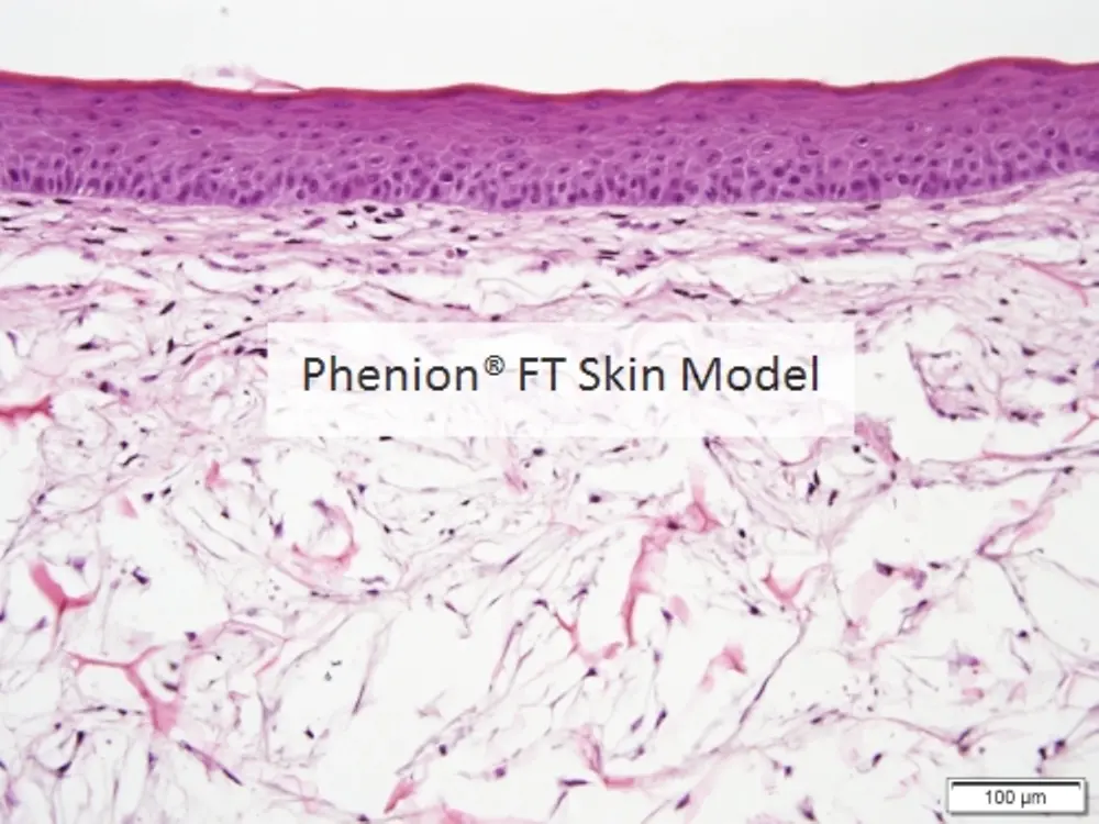
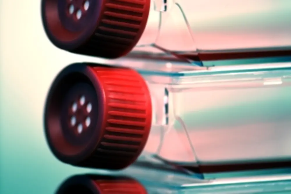
The Phenion FT Skin model is ideally suited to study the interactions between epidermis and dermis. Based on our special culture conditions, the skin model differentiates and stratifies similar to native human skin, which includes the generation of a horny layer. Analyses can be conducted with the whole tissue as well as with both compartments, dermis and epidermis, separately. A protocol for the enzymatic separation of the tissues can be found in our download center, together with protocols for the extraction and analysis of DNA, RNA and proteins from the skin tissue.
Order Details:
The Phenion® Full-Thickness (FT) Skin Model can be ordered in 3 different versions, the FT Standard version, the FT INSERT version, and the FT LARGE version.
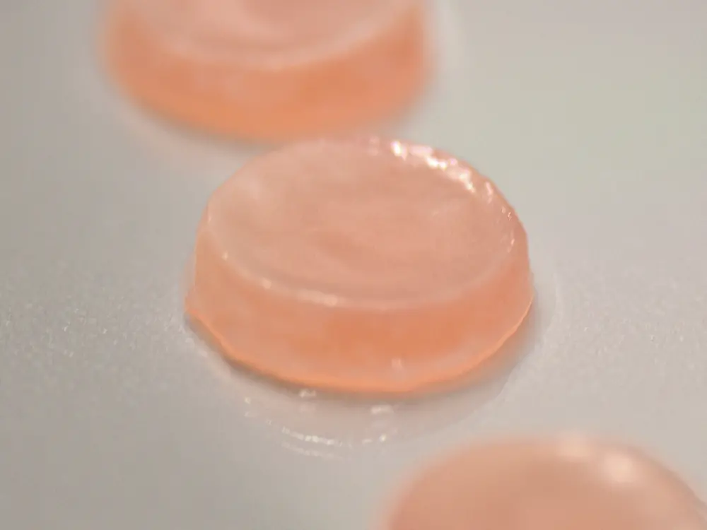
The Phenion® FT Skin Model is characterized by a diameter of 1.4 cm, which results in a tissue surface area of 1.5 cm2.
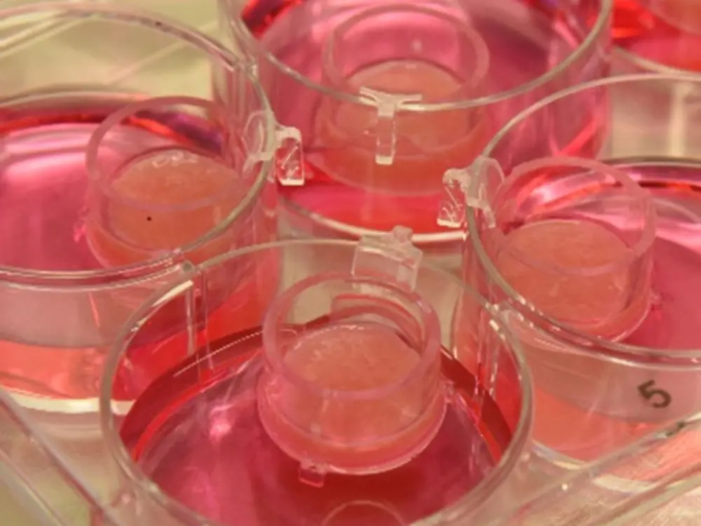
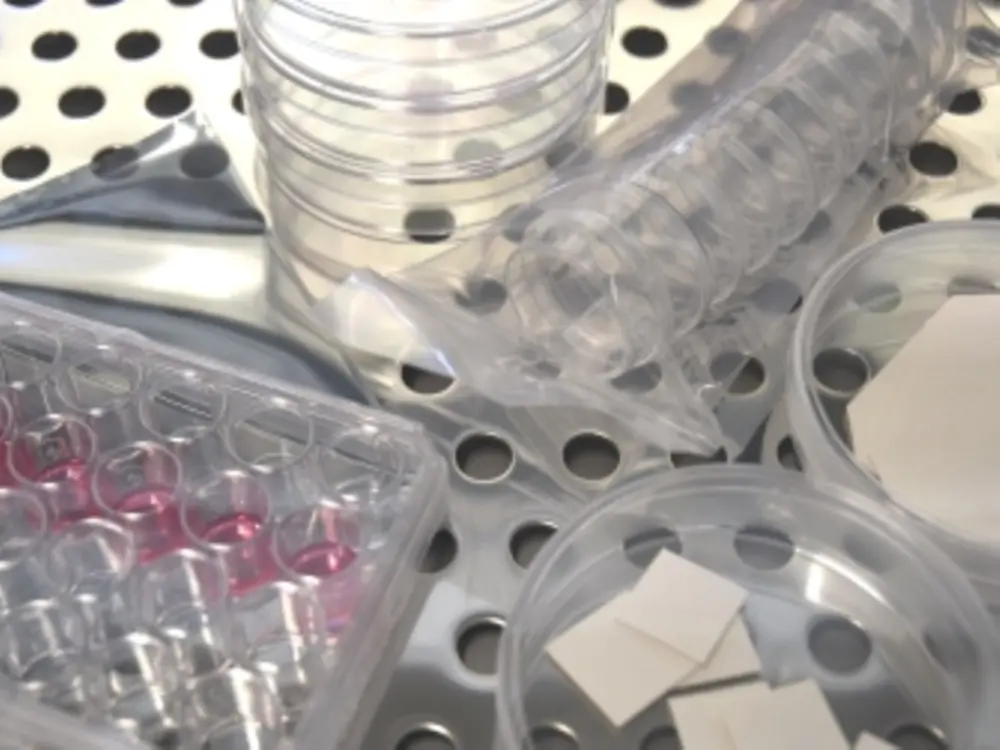
The Phenion FT Skin Model can be purchased at any number (order number FT SM-1). Together with the Phenion FT Skin Models sufficient cell culture material, required for culturing the tissues in your lab (sterile petri dishes, filter papers, spacers) is provided. These consumables enable either individual tissue culture in separate vessels (small petri dishes) or the joint culture of up to 6 models in a single medium-sized petri dish.
Tailor-made culture medium needs to be ordered separately. We recommend fresh medium for proper culture conditions every other day, the respective medium volume should be calculated with 5 ml per tissue. You will find more details in the Instructions for Use.
The FT INSERT version:
The Phenion® FT INSERT Skin Model has the same size as the FT standard skin model (surface area 1.5 cm2, diameter 1.4 cm), but is produced in co-culture inserts on a polyethylene membrane. Tissue architecture and physiological properties are identical to the standard version.
The defined insert compartment above the tissue surface allows e.g. the inoculation with microorganisms without contaminating the rest of the culture system, as well as the treatment with larger volumes of test items. This format also paves the way for the automation of tests, as the tissues in inserts can be precisely handled by robotic elements.
The specially designed inserts allow the culture of the tissues either in a hanging position, preferably in 6-well or 12-well plates, or, due to small sockets underneath the insert, in an upright standing position in culture vessels of any format. With this flexibility in use the fields of application for the Phenion® FT Skin Model are further expanded.
The FT INSERT models (order number FT INSERT-1) can be purchased at any number. Sufficient cell culture material (sterile 6-well plates) required for culturing the tissues in your lab is provided with each shipment.
Tailor-made culture medium needs to be ordered separately. We recommend fresh medium for proper culture conditions every other day, the respective medium volume should be calculated with 5 ml for hanging culture and 2 ml for standing inserts in a 6-well plate. Alternatively, the tissues can be maintained in a 12-well plate, too, which requires 2.2 ml culture medium in the hanging and 0.8 ml in the standing position. You will find more details in the Instructions for Use.
The Phenion® FT LARGE Skin Model features a tissue surface area of 7.7 cm2 which corresponds to a diameter of 3.1 cm. Tissue architecture and physiological properties are identical to the standard version.
The minimal order quantity comprises 2 FT LARGE models (order number FT LARGE-1). Higher skin model quantities can be purchased according to your individual needs. All Phenion FT LARGE Model shipments are equipped with sufficient cell culture material required for culturing the tissues in your lab (sterile petri dishes, filter papers, spacers). The material delivered with the LARGE models enables either individual tissue culture in separate vessels or the joint culture of 2 models each.
Tailor-made culture medium needs to be ordered separately. We recommend fresh medium for proper culture conditions every other day, the respective medium volume should be calculated with 35 ml/large petri dish (∅ 100 mm).
Please be aware that you also need to order a respective volume of Air-Liquid interface culture medium (order number ALI-CM-250) to take the FT models back into culture after arrival in your lab. The ALI medium has been designed to best support viability, tissue architecture and physiology of the Full-Thickness Skin models. As an indicator, a volume of 250 ml ALI medium is sufficient to culture 6 standard FT models in separate petri dishes for up to 10 days.
We provide the Air-Liquid Interface culture medium in different compositions, e.g. without phenol red, hydrocortisone or antibiotics, thus meeting your individual experimental strategies.
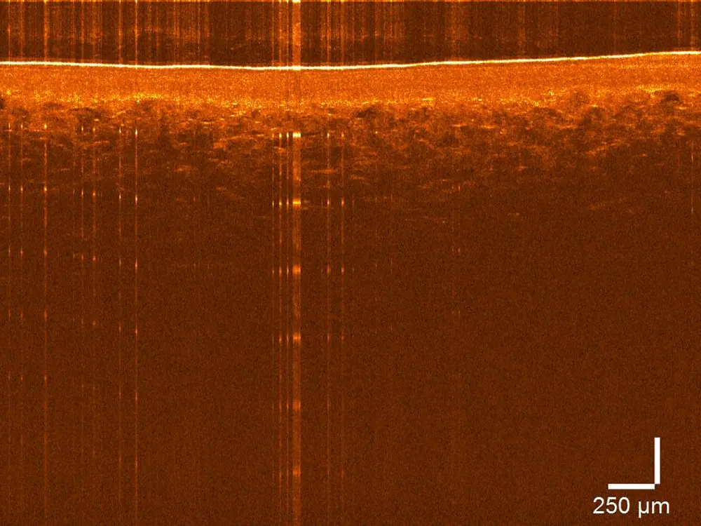
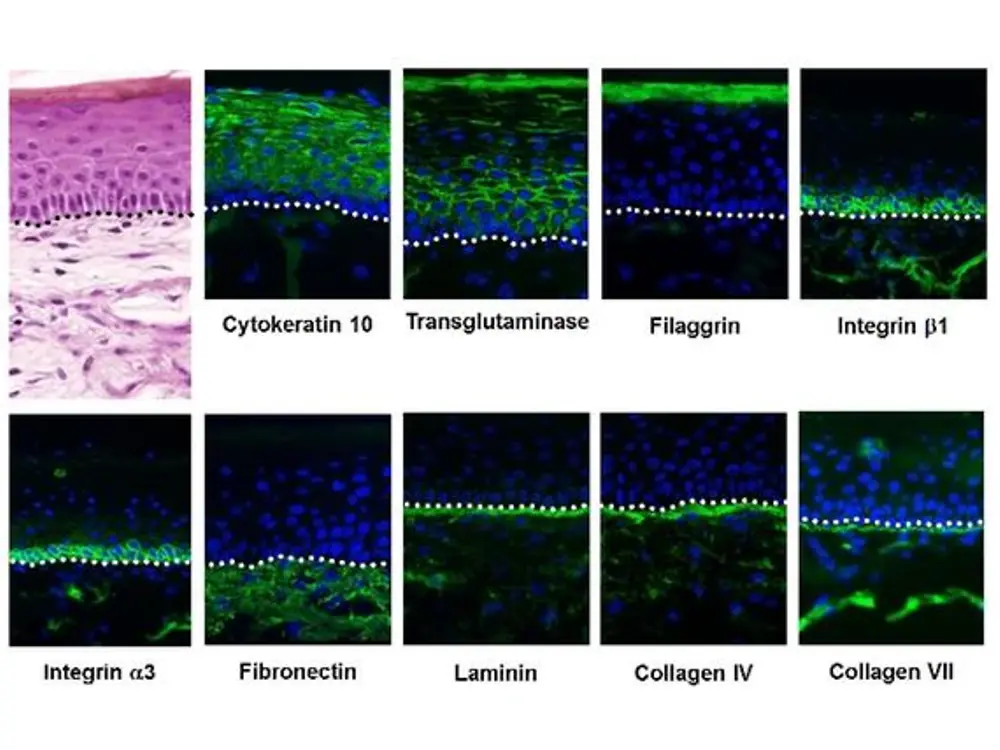
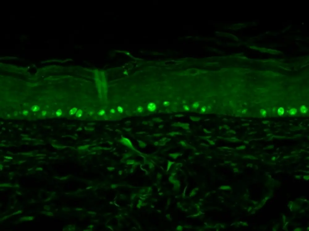
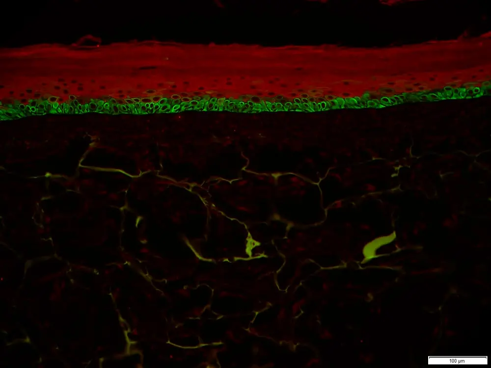
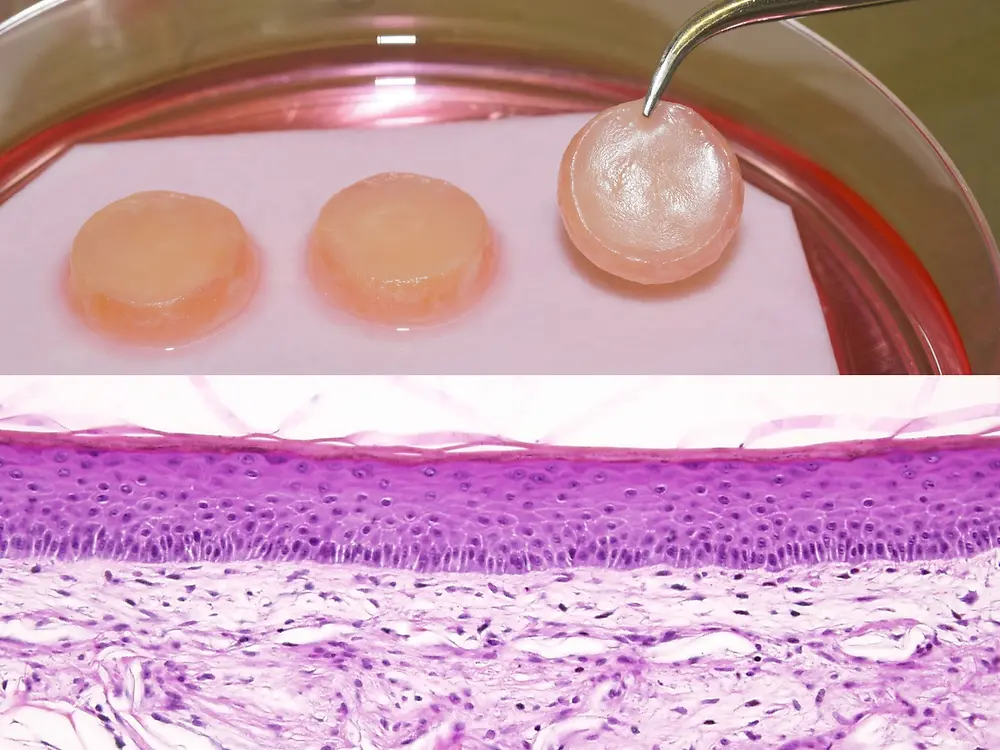
The human Phenion Full-Thickness Skin Model mimics native human skin in diverse dimensions, such as in histological architecture and in a wide spectrum of physiological and biochemical parameters. Its biological equivalence with human skin makes the tissue perfectly suited as in vitro alternative to animal testings, e.g. in the 3D Skin Comet assay for genotoxicity testing and in a vast variety of research applications as listed below.
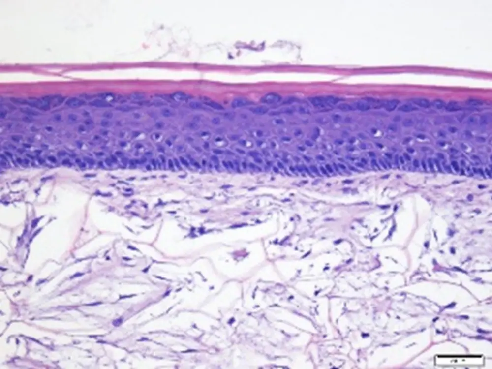
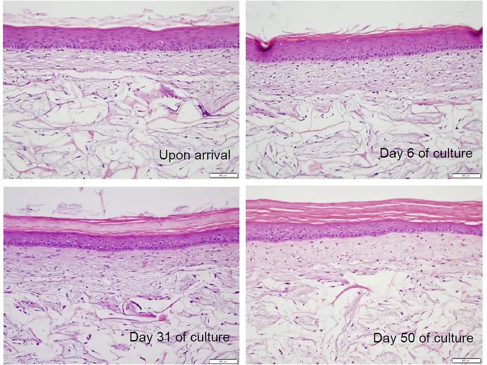
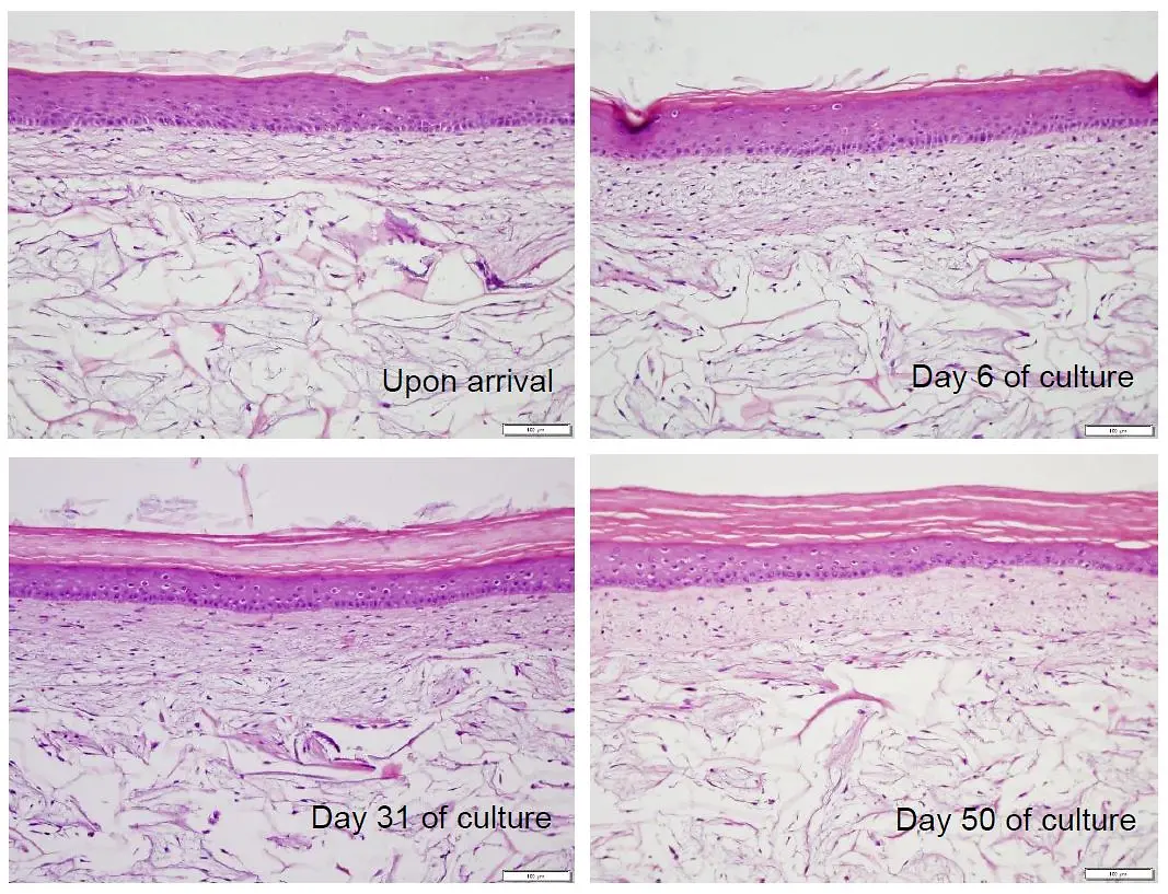
Phenion FT LL, tissue morphology (H&E stain, paraffin sections)
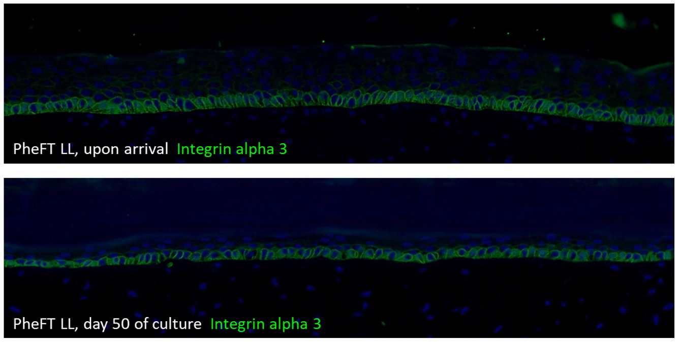
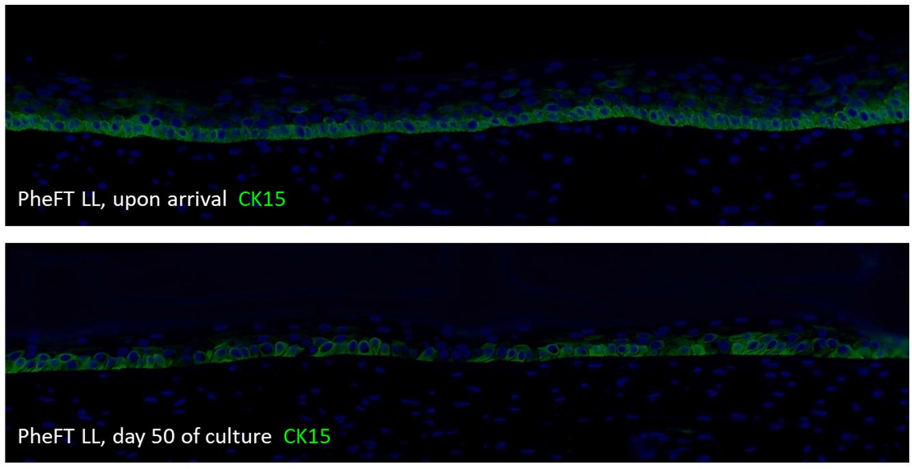
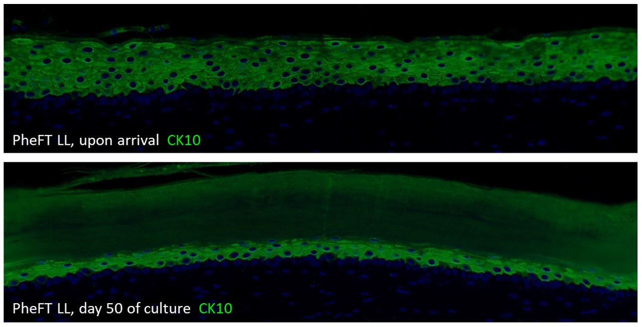
With the Phenion FT LONG-LIFE model we are offering a full-thickness skin equivalent which can be kept in culture for up to 50 days after arrival at your lab. Based on innovative protocols, fibroblasts and keratinocytes have achieved an advanced state of homeostasis at a very early stage during tissue production, which is maintained throughout the whole culture period. The 4 times extended life-span, compared to the standard FT model, opens the gateway to completely new application fields.
It allows to monitor physiological, biochemical and genetic processes, e.g. after tissue exposure with chemicals, formulations or environmental impacts, over a long period of time. Late-onset or long-term effects can be studied as well as recovery or regeneration after tissue treatment. The LONG-LIFE model is also ideally suited for repeated dosing, and it could provide an in vitro platform for research into development and treatment of skin tumors.
The standardized LONG-LIFE production processes guarantee the same high level of quality offered by the other Phenion skin models.
The Phenion FT LONG-LIFE model can be purchased at any number (order number FT LONG-LIFE-1) at a price of 228 $ per piece. Together with the Phenion FT LONG-LIFE Skin Models we provide sterile petri dishes, filter papers, spacers which are required for culturing the tissues in your lab. This material enables either individual tissue culture in separate vessels (small petri dishes) or the joint culture of up to 6 models in a single medium-sized petri dish.
Tailor-made culture medium needs to be ordered separately. We recommend fresh medium for proper culture conditions every other day, the respective medium volume should be calculated with 5 ml per tissue. You will find more details in the Instructions for Use provided on our website phenion.com.
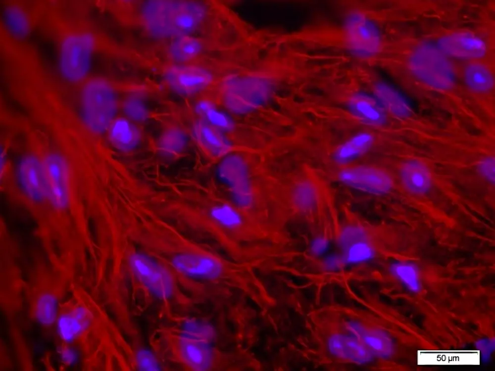
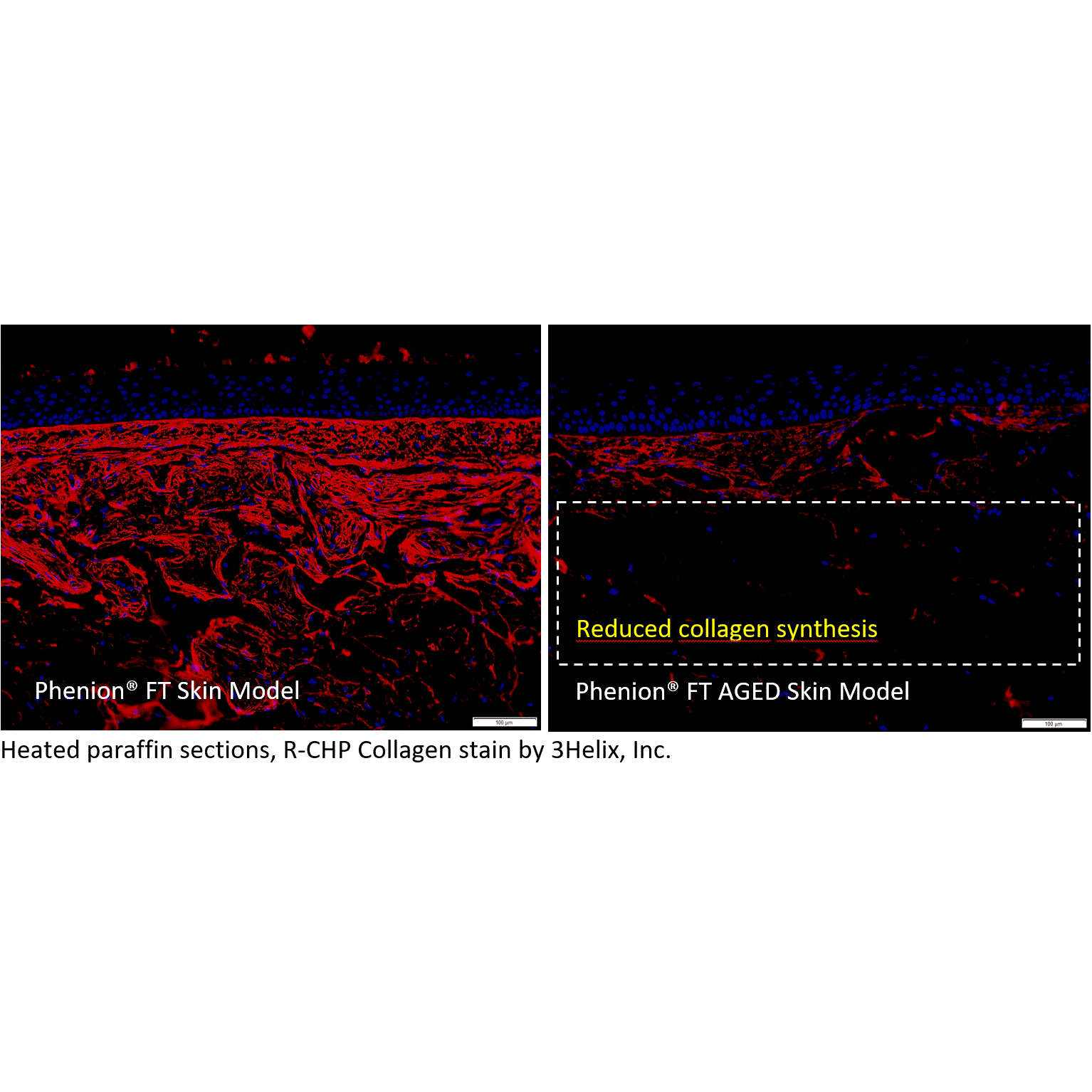
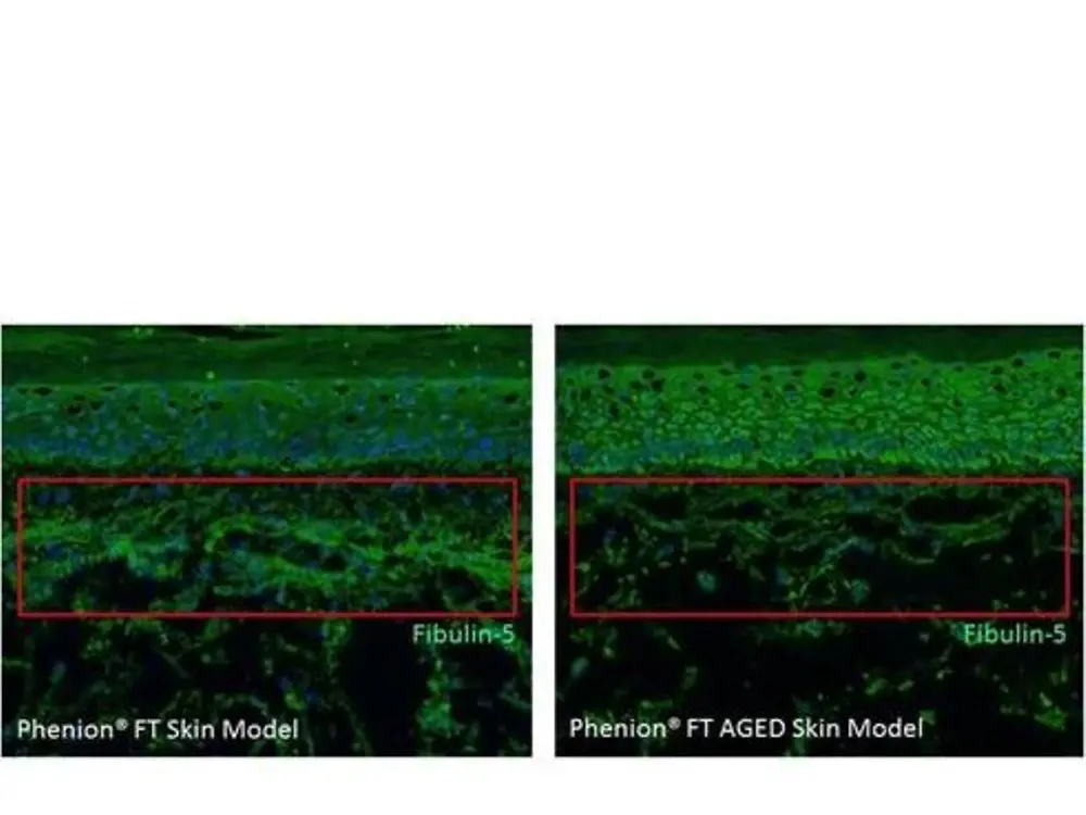
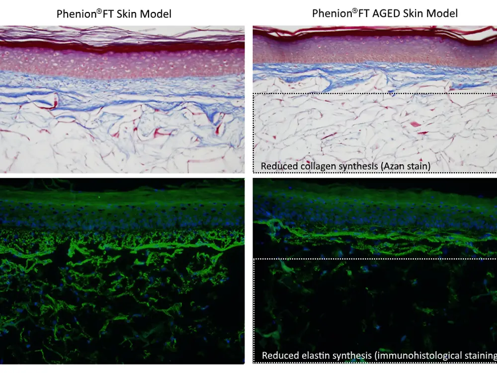
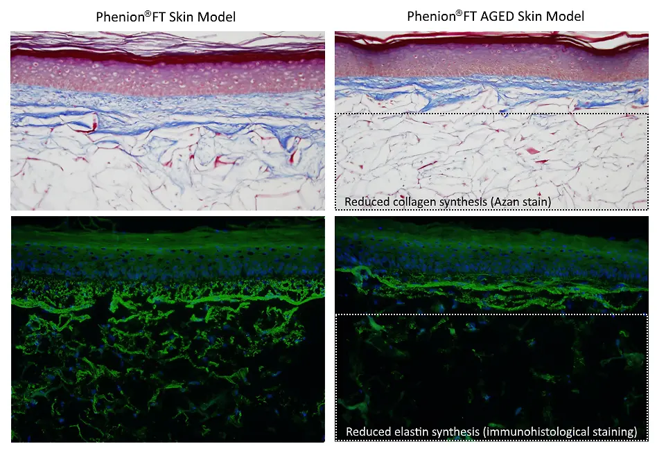
The Phenion FT AGED Skin Model is a surrogate for aged human skin. It mimics mature human skin, both in structure and physiology. The Phenion FT AGED Skin Model is characterized by a connective tissue with senescent fibroblasts, reduced synthesis of extracellular matrix (ECM) proteins like collagen and elastin, and elevated MMP secretion (Scholz, 2014, Master thesis; Dieckmann et al., 2016). With these features, the Phenion FT AGED Skin Model is an ideal research tool to study the basic biology of skin ageing mechanisms and to develop formulations which can delay or even counteract ageing.
In the Phenion FT AGED Skin Model the dermal fibroblasts are treated with mitomycin C (MMC) for 48 hours before adding the keratinocytes. The cytostatic drug MMC is an alkylating agent, which, after becoming activated within the cells, leads to covalent crosslinking of the DNA strands (Lee et al., 2006). Crosslinked DNA sections cannot be further transcribed or replicated, and cell proliferation stops. In addition, activated MMC generates reactive oxygen species (ROS), which can damage the DNA, too (Alili et al., 2014). Both effects, cell cycle arrest and ROS generation, are characteristic for the extrinsic and intrinsic ageing of the human skin. It could be demonstrated that human dermal fibroblasts can be arrested in the cell cycle and driven into senescence when they are treated with MMC in culture.
Features
Phenion® AGED Skin Model
Reduced proliferation of Fibroblasts (BrdU-Assay)
Increased ROS (2’,7’-Dichlorfluorescein-Assay)
Increased MMP-1 activity (ELISA-Assay)
Reduced gene expression for Collagen type-1 and -3 (RT-PCR)
Reduced collagen synthesis (Procollagen Type 1 C-peptide ELISA)
Reduced gene expression for Elastin (RT-PCR)
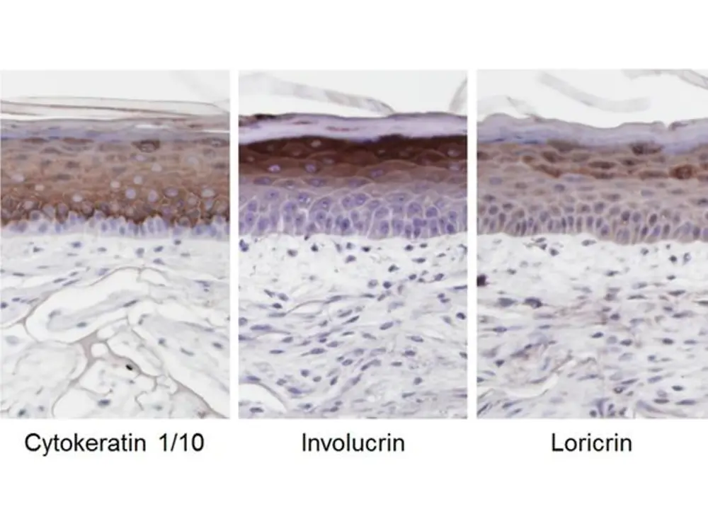
Reduced synthesis of Elastin, Filaggrin, Loricrin, Involucrin (Immunohistochemistry)
The table exemplifies some skin ageing features of the Phenion® AGED Skin Model in comparison to our standard full thickness skin model.
The minimal order quantity comprises 6 FT AGED skin models (Phenion FT AGED Skin Model Kit, order number FT AGED-6) and can be purchased at a price of 1080 $/kit.
Please note: kits with higher skin model quantities can be purchased at a price of 180 $ per additional tissue, according to your individual needs. The Phenion FT AGED Skin Model Kits are equipped with sufficient materials required for the culture of the tissues. The materials delivered with the kits enable individual tissue culture in six separate vessels or the joint cultivation (e.g. 2 x 3 or 1 x 6) of the skin models.
Content of the Phenion FT AGED Skin Model kit*
6
Full Thickness Skin Models (placed on transport-agar in a 24-well plate)
6
Sterile filter spacers
6
Sterile small petri dishes (35/10 mm)
2
Sterile large petri dishes (100/20 mm)
6
Sterile pieces of filter paper (approx. 20 x 20 mm)
1
Sterile piece of filter paper (approx. 55 x 65 mm)
*Please order 250 ml ALI-Medium (ready-to-use) separately to culture six Skin Models.
Phenion FT Skin Model Kit Instructions for Use
For further instructions, please take note of the Phenion FT Skin Model Kit Instructions for Use, available here.
The Phenion FT Skin model is ideally suited to study the interactions between epidermis and dermis. Based on our special culture conditions, the skin model differentiates and stratifies similar to native human skin, which includes the generation of a horny layer. Analyses can be conducted with the whole tissue as well as with both compartments, dermis and epidermis, separately. A protocol for the enzymatic separation of the tissues can be found in our download center, together with protocols for the extraction and analysis of DNA, RNA and proteins from the skin tissue.
Order Details:
The Phenion® Full-Thickness (FT) Skin Model can be ordered in 3 different versions, the FT Standard version, the FT INSERT version, and the FT LARGE version.
The Phenion® FT Skin Model is characterized by a diameter of 1.4 cm, which results in a tissue surface area of 1.5 cm2.
The Phenion FT Skin Model can be purchased at any number (order number FT SM-1). Together with the Phenion FT Skin Models sufficient cell culture material, required for culturing the tissues in your lab (sterile petri dishes, filter papers, spacers) is provided. These consumables enable either individual tissue culture in separate vessels (small petri dishes) or the joint culture of up to 6 models in a single medium-sized petri dish.
Tailor-made culture medium needs to be ordered separately. We recommend fresh medium for proper culture conditions every other day, the respective medium volume should be calculated with 5 ml per tissue. You will find more details in the Instructions for Use.
The FT INSERT version:
The Phenion® FT INSERT Skin Model has the same size as the FT standard skin model (surface area 1.5 cm2, diameter 1.4 cm), but is produced in co-culture inserts on a polyethylene membrane. Tissue architecture and physiological properties are identical to the standard version.
The defined insert compartment above the tissue surface allows e.g. the inoculation with microorganisms without contaminating the rest of the culture system, as well as the treatment with larger volumes of test items. This format also paves the way for the automation of tests, as the tissues in inserts can be precisely handled by robotic elements.
The specially designed inserts allow the culture of the tissues either in a hanging position, preferably in 6-well or 12-well plates, or, due to small sockets underneath the insert, in an upright standing position in culture vessels of any format. With this flexibility in use the fields of application for the Phenion® FT Skin Model are further expanded.
The FT INSERT models (order number FT INSERT-1) can be purchased at any number. Sufficient cell culture material (sterile 6-well plates) required for culturing the tissues in your lab is provided with each shipment.
Tailor-made culture medium needs to be ordered separately. We recommend fresh medium for proper culture conditions every other day, the respective medium volume should be calculated with 5 ml for hanging culture and 2 ml for standing inserts in a 6-well plate. Alternatively, the tissues can be maintained in a 12-well plate, too, which requires 2.2 ml culture medium in the hanging and 0.8 ml in the standing position. You will find more details in the Instructions for Use.
The Phenion® FT LARGE Skin Model features a tissue surface area of 7.7 cm2 which corresponds to a diameter of 3.1 cm. Tissue architecture and physiological properties are identical to the standard version.
The minimal order quantity comprises 2 FT LARGE models (order number FT LARGE-1). Higher skin model quantities can be purchased according to your individual needs. All Phenion FT LARGE Model shipments are equipped with sufficient cell culture material required for culturing the tissues in your lab (sterile petri dishes, filter papers, spacers). The material delivered with the LARGE models enables either individual tissue culture in separate vessels or the joint culture of 2 models each.
Tailor-made culture medium needs to be ordered separately. We recommend fresh medium for proper culture conditions every other day, the respective medium volume should be calculated with 35 ml/large petri dish (∅ 100 mm).
Please be aware that you also need to order a respective volume of Air-Liquid interface culture medium (order number ALI-CM-250) to take the FT models back into culture after arrival in your lab. The ALI medium has been designed to best support viability, tissue architecture and physiology of the Full-Thickness Skin models. As an indicator, a volume of 250 ml ALI medium is sufficient to culture 6 standard FT models in separate petri dishes for up to 10 days.
We provide the Air-Liquid Interface culture medium in different compositions, e.g. without phenol red, hydrocortisone or antibiotics, thus meeting your individual experimental strategies.
The human Phenion Full-Thickness Skin Model mimics native human skin in diverse dimensions, such as in histological architecture and in a wide spectrum of physiological and biochemical parameters. Its biological equivalence with human skin makes the tissue perfectly suited as in vitro alternative to animal testings, e.g. in the 3D Skin Comet assay for genotoxicity testing and in a vast variety of research applications as listed below.
Phenion FT LL, tissue morphology (H&E stain, paraffin sections)
With the Phenion FT LONG-LIFE model we are offering a full-thickness skin equivalent which can be kept in culture for up to 50 days after arrival at your lab. Based on innovative protocols, fibroblasts and keratinocytes have achieved an advanced state of homeostasis at a very early stage during tissue production, which is maintained throughout the whole culture period. The 4 times extended life-span, compared to the standard FT model, opens the gateway to completely new application fields.
It allows to monitor physiological, biochemical and genetic processes, e.g. after tissue exposure with chemicals, formulations or environmental impacts, over a long period of time. Late-onset or long-term effects can be studied as well as recovery or regeneration after tissue treatment. The LONG-LIFE model is also ideally suited for repeated dosing, and it could provide an in vitro platform for research into development and treatment of skin tumors.
The standardized LONG-LIFE production processes guarantee the same high level of quality offered by the other Phenion skin models.
The Phenion FT LONG-LIFE model can be purchased at any number (order number FT LONG-LIFE-1) at a price of 228 $ per piece. Together with the Phenion FT LONG-LIFE Skin Models we provide sterile petri dishes, filter papers, spacers which are required for culturing the tissues in your lab. This material enables either individual tissue culture in separate vessels (small petri dishes) or the joint culture of up to 6 models in a single medium-sized petri dish.
Tailor-made culture medium needs to be ordered separately. We recommend fresh medium for proper culture conditions every other day, the respective medium volume should be calculated with 5 ml per tissue. You will find more details in the Instructions for Use provided on our website phenion.com.
The Phenion FT AGED Skin Model is a surrogate for aged human skin. It mimics mature human skin, both in structure and physiology. The Phenion FT AGED Skin Model is characterized by a connective tissue with senescent fibroblasts, reduced synthesis of extracellular matrix (ECM) proteins like collagen and elastin, and elevated MMP secretion (Scholz, 2014, Master thesis; Dieckmann et al., 2016). With these features, the Phenion FT AGED Skin Model is an ideal research tool to study the basic biology of skin ageing mechanisms and to develop formulations which can delay or even counteract ageing.
In the Phenion FT AGED Skin Model the dermal fibroblasts are treated with mitomycin C (MMC) for 48 hours before adding the keratinocytes. The cytostatic drug MMC is an alkylating agent, which, after becoming activated within the cells, leads to covalent crosslinking of the DNA strands (Lee et al., 2006). Crosslinked DNA sections cannot be further transcribed or replicated, and cell proliferation stops. In addition, activated MMC generates reactive oxygen species (ROS), which can damage the DNA, too (Alili et al., 2014). Both effects, cell cycle arrest and ROS generation, are characteristic for the extrinsic and intrinsic ageing of the human skin. It could be demonstrated that human dermal fibroblasts can be arrested in the cell cycle and driven into senescence when they are treated with MMC in culture.
Features
Phenion® AGED Skin Model
Reduced proliferation of Fibroblasts (BrdU-Assay)
Increased ROS (2’,7’-Dichlorfluorescein-Assay)
Increased MMP-1 activity (ELISA-Assay)
Reduced gene expression for Collagen type-1 and -3 (RT-PCR)
Reduced collagen synthesis (Procollagen Type 1 C-peptide ELISA)
Reduced gene expression for Elastin (RT-PCR)
Reduced synthesis of Elastin, Filaggrin, Loricrin, Involucrin (Immunohistochemistry)
The table exemplifies some skin ageing features of the Phenion® AGED Skin Model in comparison to our standard full thickness skin model.
The minimal order quantity comprises 6 FT AGED skin models (Phenion FT AGED Skin Model Kit, order number FT AGED-6) and can be purchased at a price of 1080 $/kit.
Please note: kits with higher skin model quantities can be purchased at a price of 180 $ per additional tissue, according to your individual needs. The Phenion FT AGED Skin Model Kits are equipped with sufficient materials required for the culture of the tissues. The materials delivered with the kits enable individual tissue culture in six separate vessels or the joint cultivation (e.g. 2 x 3 or 1 x 6) of the skin models.
Content of the Phenion FT AGED Skin Model kit*
6
Full Thickness Skin Models (placed on transport-agar in a 24-well plate)
6
Sterile filter spacers
6
Sterile small petri dishes (35/10 mm)
2
Sterile large petri dishes (100/20 mm)
6
Sterile pieces of filter paper (approx. 20 x 20 mm)
1
Sterile piece of filter paper (approx. 55 x 65 mm)
*Please order 250 ml ALI-Medium (ready-to-use) separately to culture six Skin Models.
Phenion FT Skin Model Kit Instructions for Use
For further instructions, please take note of the Phenion FT Skin Model Kit Instructions for Use, available here.